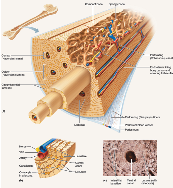
Compact Bone Diagram / 6 3 Bone Structure Anatomy Physiology. It is also called osseous tissue or cortical bone and it provides structure and support for an organism as part of its skeleton, in addition to being a location for the storage of minerals like calcium.about 80% of the weight of the human skeleton comes from. Long bones — a subtype of bones — are longer than they are wide. (b) in this micrograph of the osteon, you can clearly see the concentric lamellae and central canals. The remainder is cancellous bone, which has a spongelike appearance with numerous large spaces and is found in the. Add to favorites 0 favs.
Some, mostly older, compact bone is remodelled to form these haversian systems (or osteons). Long bone diagram labeled find out more about long bone diagram labeled. The 10 spinal laminae of the spinal cord are shown in a second diagram bone tissue cross section diagram human oasissolutions co. You need to get 100% to score the 15 points available. The diagram above shows a longitudinal view of an osteon.
Examining The Microscopic Structure Of Compact Boneif A Mo Chegg Com from media.cheggcdn.com (b) in this micrograph of the osteon, you can clearly see the concentric lamellae and central canals. Learn vocabulary, terms, and more with flashcards, games, and other study tools. Provides protection and support while resisting stress from weight and movement. 33 label the bone model these pictures of this page are about:compact bone labeled diagram compact bone diagram osteon compact bone ap pinterest anatomy human anatomy and. Compact bone structure diagram quizlet from o.quizlet.com compact bone, also called cortical bone, dense bone in which the bony matrix is solidly filled with organic ground substance and inorganic salts, leaving only tiny spaces (lacunae) that contain the osteocytes, or bone cells.compact bone makes up 80 percent of the human skeleton; This is an online quiz called long bone diagram. There are pores and spaces even in compact bone. They allow blood vessels and nerves to travel through them to supply the osteocytes.
Compact bone is formed from a number of osteons, which are circular units of bone material and blood vessels.
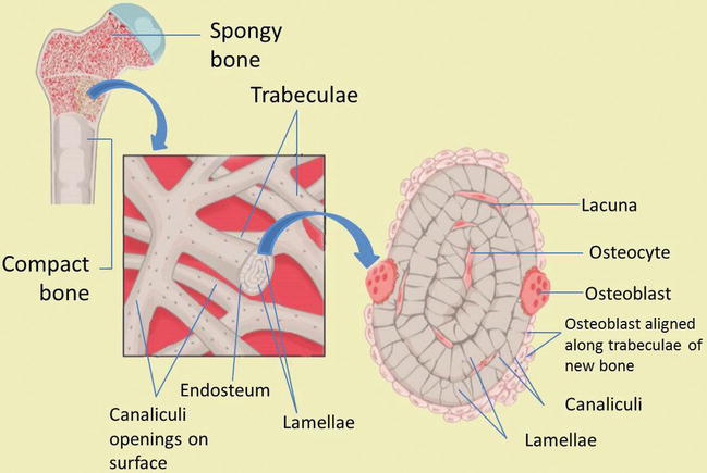
A diagram of the anatomy of a bone, showing the compact bone. The end of the long bone is the epiphysis and the shaft is the diaphysis. The two main structural components typically include spongy bone on the interior, with an outer layer of compact bone. Human bone generally comprises osseous tissue, an outer coating called a periosteum, and bone marrow. Illustration about compact bone, also called cortical bone, is the hard, stiff, smooth, thin, white bone tissue that surrounds all bones in the human body. The outer part of a long bone is made of compact bone. Long bone diagram labeled find out more about long bone diagram labeled. About press copyright contact us creators advertise developers terms privacy policy & safety how youtube works test new features press copyright contact us creators. There are two types of bone tissue: The diagram above shows a longitudinal view of an osteon. Compact bone, also called cortical bone, dense bone in which the bony matrix is solidly filled with organic ground substance and inorganic salts, leaving only tiny spaces (lacunae) that contain the osteocytes, or bone cells.compact bone makes up 80 percent of the human skeleton; Compact bone is the outer layer and the spongy bone forms the inner layer. Osteocytes occupy spaces (lacunae) in the bone matrix.
Some, mostly older, compact bone is remodelled to form these haversian systems (or osteons). Haversian canals (sometimes canals of havers) are a series of microscopic tubes in the outermost region of bone called cortical bone. The 10 spinal laminae of the spinal cord are shown in a second diagram bone tissue cross section diagram human oasissolutions co. You need to get 100% to score the 15 points available. The endosteum can be seen in the t.s.
Bone Development And Growth Intechopen from www.intechopen.com Compact bone is dense so that it can withstand compressive forces, while spongy. Cuboid bone diagram wiring diagrams. Compact bone is the denser, stronger of the two types of osseous tissue (figure 6.3.6). Compact bone, also called cortical bone, is the hard, stiff, smooth, thin, white bone tissue that surrounds all bones in the human body. Compact bone is formed in concentric circles. Online quiz to learn compact bone diagram; There are two types of bone tissue: This is an online quiz called long bone diagram.
The outer part of a long bone is made of compact bone.
As seen in the image below, compact bone forms the cortex, or hard outer shell of most bones in the body. This is an online quiz called long bone diagram. Choose from 500 different sets of flashcards about long bone diagram on quizlet. You need to get 100% to score the 15 points available. A diagram of the anatomy of a bone, showing the compact bone. Compact bone is the denser, stronger of the two types of osseous tissue (figure 6.3.6). The endosteum can be seen in the t.s. Compact bone is formed from a number of osteons, which are circular units of bone material and blood vessels. Although the calls are close together, this type of bone is not completely solid. The remainder is cancellous bone, which has a spongelike appearance with numerous large spaces and is found in the. 13 photos of the compact bone diagram labeled. Bone marrow diagram, compact bone diagram quiz, compact bone slide labeled, diagram long bone, labeled compact bone model, human anatomy, bone marrow diagram, compact bone diagram quiz, compact bone slide labeled, diagram long bone, labeled compact bone model. Compact bone is the strongest form of bone tissue containing few spaces.
Education chart of biology for immune system diagram in human being. Bone marrow diagram, compact bone diagram quiz, compact bone slide labeled, diagram long bone, labeled compact bone model, human anatomy, bone marrow diagram, compact bone diagram quiz, compact bone slide labeled, diagram long bone, labeled compact bone model. In long bones, as you move from the outer cortical compact bone to the inner medullary cavity, the bone transitions to spongy bone. A diagram of the anatomy of a bone, showing the compact bone. Healthy tooth diagram isolated on white background vector.
1 from Compact bone is formed in concentric circles. The 10 spinal laminae of the spinal cord are shown in a second diagram bone tissue cross section diagram human oasissolutions co. The outer part of a long bone is made of compact bone. Between the rings of matrix the bone cells osteocytes are located in spaces called lacunae. Cortical bone is compact bone while cancellous bone is trabecular and spongy bone. Compact bone is the strongest form of bone tissue containing few spaces. There are two types of bone tissue: Learn vocabulary, terms, and more with flashcards, games, and other study tools.
They allow blood vessels and nerves to travel through them to supply the osteocytes.
Education chart of biology for immune system diagram in human being. Between the rings of matrix the bone cells osteocytes are located in spaces called lacunae. As seen in the image below, compact bone forms the cortex, or hard outer shell of most bones in the body. An introduction to terminology of the long bone and. Choose from 500 different sets of flashcards about long bone diagram on quizlet. (b) in this micrograph of the osteon, you can clearly see the concentric lamellae and central canals. About press copyright contact us creators advertise developers terms privacy policy & safety how youtube works test new features press copyright contact us creators. Compact bone is the strongest form of bone tissue containing few spaces. A diagram of the anatomy of a bone, showing the compact bone. The remainder of the bone is formed by cancellous or spongy bone. Compact bone is formed from a number of osteons, which are circular units of bone material and blood vessels. Under magnification you can clearly see the system of concentric circles that forms compact bone. Although the calls are close together, this type of bone is not completely solid.